
The Inscopix nVue™ system that was released last year has been a game changer for the in-vivo freely behaving imaging field and we are excited about the new application that enables simultaneous imaging of blood flow with cellular activity! If you aren’t familiar, the nVue system is a miniaturized microscope enabling the dual color imaging of two distinct brain signals- neural activity and blood flow- in the green and red channel respectively in freely behaving subjects, complete with a data acquisition and analysis software and world class support.
To introduce myself, my name is Srishti Gulati, and I am an Application Scientist at Inscopix. I lead new application development and validation using Inscopix products and I am beyond thrilled to be a part of this brand-new application enabling simultaneous imaging of blood flow and calcium signals with the nVue system! I hope that this blog post gives you a fun behind-the-scenes look at the development and release of this impactful new miniscope application.
The goal of this application is to accurately track red blood cells (RBCs) flowing through thin vessels while measuring neuronal calcium activity. For this, the in-vivo data is required to be collected at a high frame rate of 100 Hz in a multiplexed manner for accurate measurement of the spatiotemporal signals. The nVue system is able to precisely control both imaging LEDs in quick succession while ensuring that there is no signal leakage between channels to avoid crosstalk. Furthermore, the unique control over the electronic focus developed by Inscopix allows for a seamless correction of the chromatic aberration that is typical of gradient refractive index (GRIN) lenses. Enabling this capability required a tremendous amount of effort from the hardware, software and UI teams that have all worked together to create an intuitive and seamless data acquisition experience as is reflected in the polished user-friendly interface.
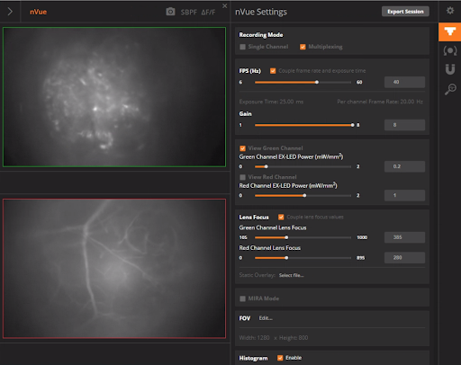
Inscopix data acquisition software UI showing a two-pane interface of green (neurons) and red (blood vessels) channels and configurable settings.
Next, labeling the animal vasculature in an awake state, to minimize any anesthesia induced artifacts, requires injection of high contrast dyes through the tail vein. Also, the dye molecules themselves are excreted out of the animal’s system within a couple of hours depending on their molecular weights. The Applications team tested blood dyes of varying molecular weights and spectral properties and identified the best candidates for application development. To validate the application, we imaged medium spiny neurons expressing GCaMP in mouse dorsal striatum along with Texas Red-dextran in the blood vessels and measured changes in activity in baseline and amphetamine conditions.
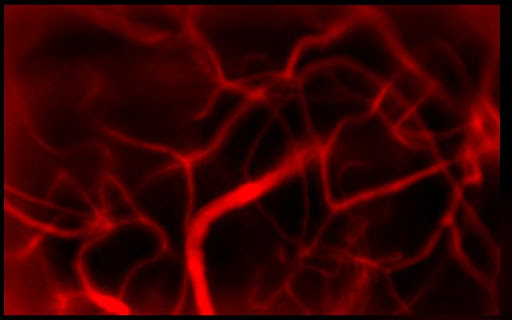
Intricate network of blood vessels in mouse dorsal striatum labeled with Texas Red-dextran as seen with an nVue system.
Parallel to the Applications team’s blood dye testing, the Data Scientists and Software Engineers were busy validating, iterating and implementing the RBC velocity algorithm into the Inscopix Data Processing Software (IDPS). The algorithm development and validation phase was an extensive process where RBCs flowing through vessels across probe types and brain regions were manually hand-annotated by scientists across thousands of frames to generate ground truth data. Data Scientists used this valuable information to develop and iterate on the algorithms to track changes in vessel diameter and RBC speed at a micron and millisecond scale. The IDPS team then took these algorithms and wrapped them in a beautiful, intuitive UI for the end user to apply on their data. This process is typical of how our applications and software teams work collaboratively to ensure a clean, robust product makes its way into the hands of our customers.
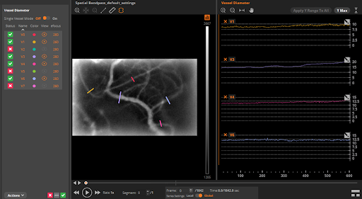
IDPS is a powerful processing software that allows researchers to gain insights into complex brain activity. Here, you can see an output of the vessel diameter function on a data set from mouse dorsal striatum.
I hope this blog provides you with an idea of what goes into a new application development at Inscopix and how we work across teams to present fully validated uses of our products. We sincerely hope that the Blood Flow Application Package will allow researchers to use miniscope platform to understand the synergistic relationship between neurons and blood flow in healthy and disease states such as stroke, addiction, Parkinson’s disease amongst various other disorders and help find ways to restore normal brain function.